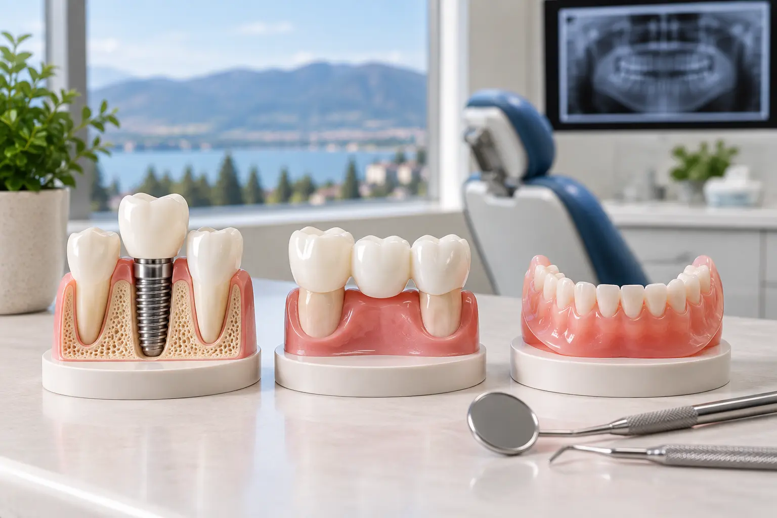

Most people think of a dental X-ray as a formality. You bite down on a small sensor, it beeps, and the appointment carries on. But if you are booked in for an implant, a root canal or a wisdom tooth extraction, that image is doing far more work than it looks like it is doing. It is the difference between a plan and a guess.
At Discover Dental, your Kelowna dentist, imaging comes first for almost every procedure more involved than a filling. Here is what we are actually looking at, why 3D imaging changed implant planning, and how much radiation is genuinely involved.
What a Digital Dental X-Ray Actually Is
Digital X-rays, or digital radiographs, use an electronic sensor instead of film. The sensor picks up the image and sends it straight to a screen. There is no chemical processing and no waiting.
The practical result is that your dentist can look at your teeth, jawbone and the structures around them in detail, and can spot things that are invisible during a normal exam. Decay between two teeth that are touching. Bone loss under the gumline. An infection sitting at the tip of a root. None of that shows up in a mirror.
Because the image is on a monitor, it can also be enlarged, contrast-adjusted and compared against images taken two years ago. That last part matters more than most patients realise, and we will come back to it.
You can read more about the equipment we use on our digital X-rays page.
The Four Types, and Why It Matters Which One You Get
“Dental X-ray” covers several different images that answer different questions. Knowing which is which helps when you are told you need one.
Bitewing
The one you get at most checkups. You bite on a small tab and it captures the crowns of the upper and lower teeth on one side. It is the best image for finding decay between teeth and for checking the fit of existing fillings and crowns.
Periapical
Shows one or two teeth from crown to root tip, including the bone around the root. This is the image used when a specific tooth is causing trouble, and it is what confirms whether an infection has reached the root.
Panoramic
A single wide image of the entire upper and lower jaw, both joints and all the teeth, including ones that have not erupted yet. Lower detail than the two above, but it shows the whole picture. Wisdom tooth assessments almost always start here.
Cone Beam CT (CBCT)
A 3D scan. Rather than a flat image, it builds a volume you can rotate and slice through in any direction. This is the one that changed implant surgery, and it is also the one that carries a meaningfully higher radiation dose than the other three, so it is used when the 3D information will actually change the treatment, not routinely.
How Much Radiation Are We Actually Talking About?
This is the question patients ask most and get the vaguest answers to, so here is a straight one.
The dose from a single digital dental X-ray is very small. The American Dental Association puts it this way: in some cases a dental X-ray delivers less radiation than you receive in a single day just from the natural background radiation around you. That background comes from the ground, from cosmic rays, and from the air, and it is happening whether you visit a dentist or not.
Digital sensors do need less exposure than the film they replaced. How much less depends on which sensor type and which film speed you compare, so we would rather give you the actual comparison above than a percentage figure that sounds impressive but does not hold up.
The principle that governs all of this is ALARA, meaning as low as reasonably achievable. In practice that means three things: only taking an image when it will change what we do, restricting the beam to the area being imaged, and using previous images wherever they answer the question. If you have had recent X-rays at another practice, tell us. We would rather request those than repeat them.
CBCT is the exception worth flagging. A 3D scan involves more radiation than a standard 2D image, which is exactly why it is reserved for cases like implant planning or a complicated extraction where the detail genuinely affects the outcome.
You can read the current guidance directly from the American Dental Association’s page on dental X-rays.
Why Accurate Imaging Matters
In dentistry, precision is everything. High-quality images are essential for planning treatments that rely on exact measurements and detailed visualization, such as dental implants, orthodontic adjustments and restorative procedures.
Digital X-rays give your dentist the clarity and accuracy needed to:
- Assess bone density and tooth positioning
- Detect structural abnormalities or hidden decay
- Evaluate root health and jaw alignment
- Identify potential complications before treatment begins
This level of accuracy means your treatment plan is built around your actual anatomy, which reduces risk and improves the result.
Digital X-Rays and Dental Implant Planning
When it comes to dental implants in Kelowna, imaging is one of the most important steps in getting a result that lasts.
An implant replaces a missing tooth with a titanium post that integrates with your jawbone and acts as an anchor for a crown or bridge. For that to work, the post has to sit in the right position with enough bone around it. Not roughly the right position. The right one.
What the Imaging Shows
- Bone health: Digital images let your dentist measure bone density and height precisely, confirming there is enough support for the implant.
- Nerve and sinus locations: Imaging identifies exactly where nerves and sinuses sit, so they can be avoided during surgery.
- Implant placement: A CBCT scan allows the implant to be placed virtually, in software, before the actual procedure. Angle, depth and position are decided in advance, which improves accuracy and shortens time in the chair.
- Healing and integration: Post-surgical images confirm the implant is fusing with the bone the way it should.
What Goes Wrong Without It
The failures that show up after implant surgery are usually not surprises. They are things that were visible beforehand and were not looked for.
Not enough bone height means the implant lacks stability and can loosen over time. Placing a post too close to the inferior alveolar nerve in the lower jaw risks lasting numbness in the lip and chin. In the upper jaw, going too far means entering the sinus. And an implant angled without reference to the teeth around it can end up with a crown that looks off or does not bite properly.
Every one of those is identifiable on a scan before anyone picks up an instrument. That is the whole argument for imaging first.
How Digital X-Rays Support Orthodontic Treatment
Imaging matters just as much in orthodontics. Whether teeth are being straightened with braces or clear aligners, accurate X-rays are what allow a precise, efficient plan.
- Tooth and jaw alignment: X-rays show how teeth sit within the jaw, including ones that have not erupted yet.
- Growth patterns: For younger patients, imaging helps predict how the jaw and facial structures will develop, which guides the timing of treatment.
- Customised planning: Digital records mean the plan is built around one patient’s anatomy rather than an average.
- Tracking progress: Follow-up images let the orthodontist monitor movement and adjust as treatment goes on.
Orthodontic treatment for our patients is handled together with our orthodontic partners, and the imaging we take here is what they work from.
Where Else X-Rays Change the Plan
Root Canal Therapy
Imaging identifies infection, maps the shape of the root canals, and confirms afterwards that every canal has been cleaned and sealed. Some teeth have more canals than expected, and a missed one is the usual reason a root canal needs redoing.
Wisdom Tooth Extraction
X-rays show where wisdom teeth sit relative to nerves, bone and the teeth beside them. That is what determines whether an extraction is straightforward or surgical, and it is decided before the appointment rather than during it. The same applies to any tooth extraction where the root shape is unusual.
Crowns, Bridges and Same-Day Restorations
For crowns and bridges, imaging guides tooth preparation and alignment, which is what produces a restoration that fits properly at the margin. Digital imaging also underpins CEREC same-day restorations, where the design and milling both happen from digital data during one visit. We covered how that process works in our post on same-day restorations.
Cracked Teeth
A crack is one of the few problems an X-ray does not always show, because a hairline fracture running vertically can be invisible on a flat image. What imaging does rule out is everything else that produces similar symptoms, and it shows whether the crack has reached the nerve or the bone. If you have a tooth that hurts intermittently, this is worth reading, and if it is painful now, contact us about emergency care.
TMJ and Bite Assessment
Detailed imaging of the jaw and joint helps diagnose alignment problems and guides treatment for jaw pain and temporomandibular disorders.
How Often Do You Actually Need X-Rays?
There is no fixed schedule, and any practice that tells you otherwise is running on habit rather than evidence. The interval depends on your risk.
Someone with no history of decay, healthy gums and good home care may go a long stretch between bitewings. Someone with active decay, gum disease, a heavy restoration history or a dry mouth needs them more often, because problems in that group move faster and the cost of catching them late is higher.
What decides it is your mouth, your history and what has changed since the last set. Age alone does not decide it, and neither does the calendar.
This is also where the ability to compare images over time earns its keep. Bone loss of half a millimetre is invisible on a single image and obvious when you put this year’s image next to the one from three years ago.
X-Rays for Children
Children are imaged less often and with the beam sized to a smaller mouth. The images serve a different purpose than they do for adults: checking that adult teeth are forming and positioned correctly, spotting decay in baby teeth that has spread further than it looks, and identifying crowding early enough that it can be planned for rather than reacted to.
If your child is anxious about the appointment itself, that is worth raising before the visit rather than during it. Our children’s dentistry page covers how we approach it, and we have written separately about when nitrous oxide is recommended for children.
What It Is Like as a Patient
The sensors are small, shaped to sit reasonably comfortably, and quick. A full set of bitewings takes a few minutes. A panoramic image takes under a minute and you stay standing, with nothing in your mouth at all.
The part most patients notice is seeing the image themselves. When a dentist points at a dark patch between two teeth on a screen in front of you, the recommendation stops being something you are asked to take on trust. If you are nervous about dental visits generally, our sedation options are worth asking about, though imaging itself is rarely the part people find difficult.
Your images are read by our dentists, who will walk you through what they show before any treatment is discussed.
Common Questions About Dental X-Rays
Are dental X-rays safe during pregnancy?
Dental imaging is considered safe during pregnancy, and delaying necessary treatment usually carries more risk than the imaging does. Tell us that you are pregnant regardless, so it can be factored into the plan.
Can I refuse a dental X-ray?
Yes. You can decline any imaging. What you should know is that some treatments cannot be done safely without it. An implant or a surgical extraction planned without imaging is a plan built on assumption, and most dentists will decline to proceed rather than take that risk.
Do I need new X-rays if I have recent ones from another dentist?
Often not. If you have had imaging elsewhere in the recent past, we can request the files. That is the preferred route, and it is written into current radiation safety guidance.
What is the difference between an X-ray and a CBCT scan?
A standard X-ray produces a flat, two-dimensional image. A CBCT produces a 3D volume that can be rotated and viewed in cross-section. CBCT gives far more information for surgical planning and involves more radiation, so it is used where the extra detail will change what happens.
Why do X-rays sometimes need to be retaken?
Usually positioning. If the sensor shifts or the angle is slightly off, part of the tooth or root can be cut off or overlapped, and an image that does not show the thing you need to see is not worth keeping. Digital imaging makes this obvious immediately rather than after processing.
Book an Appointment at Discover Dental
If you are considering an implant, have a tooth that needs assessing, or are simply due for a checkup, we will talk you through what the imaging shows before we discuss any treatment.
Discover Dental is at 3975 Lakeshore Rd #301, Kelowna, BC V1W 1V3, open Monday to Friday, 8:00 AM to 5:00 PM. Call (778) 477-5554 or request an appointment online.